Abdominal Anatomy / Internal Anatomy Of Male Chest And Abdomen On White Stock Photo - Download Image Now - iStock. Respiratory muscle training online course: Abdominal anatomy, abdomen, gastrointestinal anatomy, gastrointestinal system. Connections between the left and the middle colic the gonadal arteries cross the abdominal ureters approximately halfway between the pelvic inlet and the renal pelvis. The abdomen (colloquially called the stomach, belly, tummy or midriff) is the part of the body between the thorax (chest) and pelvis, in humans and in other vertebrates. The above lines intersect and divide the abdomen into nine regions (clockwise from the top)
Lee moffitt cancer center & research institute in. This page provides a photo gallery that presents the anatomy of the abdomen by means of ct (axial, coronal, and sagittal reconstructions). Level of l5, near transtubercular plane anatomy ileum, rectus abdominis muscle, ileocecal junction, cecum, internal abdominal oblique muscle, external abdominal oblique muscle, psoas major muscle, iliacus muscle, body of l5 vertebra. Respiratory muscle training strengthen the function of the respiratory. This section of the website will explain large and minute details of abdomen axial cross sectional anatomy.
Pelvic stress fracture: a rare but real risk in distance athletes from sportsinjury.wpengine.com This page provides a photo gallery that presents the anatomy of the abdomen by means of ct (axial, coronal, and sagittal reconstructions). The above lines intersect and divide the abdomen into nine regions (clockwise from the top) Level of l5, near transtubercular plane anatomy ileum, rectus abdominis muscle, ileocecal junction, cecum, internal abdominal oblique muscle, external abdominal oblique muscle, psoas major muscle, iliacus muscle, body of l5 vertebra. Two layers in abdomenfatty superficial layer (camper's fascia)deeper membranous layer (scarper's fascia). The abdomen contains many vital organs: 5 name the nine abdominal regions and their main contents. Related online courses on physioplus. Sciency root words make anatomical parts harder to memorize.
The abdominal wall is the wall enclosing the abdominal cavity that holds a bulk of gastrointestinal viscera.
Simple, easy notes for quick revision of important questions. Radiology basics of abdominal ct anatomy with annotated coronal images and scrollable axial images to help medical students and junior doctors learning anatomy. Abdominal anatomy, abdomen, gastrointestinal anatomy, gastrointestinal system. Abdominal anatomy seen on ct. The abdomen refers to the region between the pelvis (pelvic brim) and the thorax (thoracic diaphragm) in vertebrates, including humans. The transversus abdominis muscle is the deepest of the abdominal muscles, lying internally to the internal abdominal obliques. Abdomen, in human anatomy, the body cavity lying between the chest or thorax above and the pelvis below and from the spine in the back to the wall the abdominal organs are supported and protected by the bones of the pelvis and ribcage and are covered by the greater omentum, a fold of peritoneum. This is a laparoscopic tour of abdominal cavity anatomy. Lee moffitt cancer center & research institute in. But with the use of smart technology, you can learn faster and master abdomen anatomy in no time! The viewer gets to see the abdominal organs just as the surgeon does while he or she is operating. Respiratory muscle training online course: Windham was previously a surgical oncologist in the sarcoma program of the h.
The above lines intersect and divide the abdomen into nine regions (clockwise from the top) The stomach, the small intestine (jejunum and ileum), the large intestine (colon), the liver, the spleen, the gallbladder, the pancreas, the uterus, the fallopian. 6 write the origin, insertion and nerve supply of muscles of anterior abdominal wall. Lee moffitt cancer center & research institute in. The xiphoid process and costal.
4: Abdomen | Basicmedical Key from basicmedicalkey.com Abdominal anatomy seen on ct. Abdominal surface anatomy can be described when viewed from in front of the abdomen in 2 ways: Abdominal wall anatomy that is clinically pertinent to the surgeon, focusing primarily on the structures of the anterior abdominal wall, will be reviewed. This mri abdomen axial cross sectional anatomy tool is absolutely free to use. The abdominal wall is the wall enclosing the abdominal cavity that holds a bulk of gastrointestinal viscera. Abdomen, in human anatomy, the body cavity lying between the chest or thorax above and the pelvis below and from the spine in the back to the wall the abdominal organs are supported and protected by the bones of the pelvis and ribcage and are covered by the greater omentum, a fold of peritoneum. The vascular anatomy distal to the middle colic artery and near the splenic flexure is variable. There are multiple anatomical areas within the abdomen, each of which contain specific contents and are bound by certain borders.
The abdomen contains many vital organs:
Abdominal anatomy, abdomen, gastrointestinal anatomy, gastrointestinal system. A good amount of area is covered by the abdominal wall. This mri abdomen axial cross sectional anatomy tool is absolutely free to use. This page provides a photo gallery that presents the anatomy of the abdomen by means of ct (axial, coronal, and sagittal reconstructions). Windham was previously a surgical oncologist in the sarcoma program of the h. The transversus abdominis muscle is the deepest of the abdominal muscles, lying internally to the internal abdominal obliques. Two layers in abdomenfatty superficial layer (camper's fascia)deeper membranous layer (scarper's fascia). These general diagrams show the digestive system, with the major human anatomical structures labeled (mouth, tongue, oral cavity, teeth, buccal glands, throat, pharynx, oesophagus, stomach, small intestine, large. There are multiple anatomical areas within the abdomen, each of which contain specific contents and are bound by certain borders. The abdomen (colloquially called the stomach, belly, tummy or midriff) is the part of the body between the thorax (chest) and pelvis, in humans and in other vertebrates. Abdomen, in human anatomy, the body cavity lying between the chest or thorax above and the pelvis below and from the spine in the back to the wall the abdominal organs are supported and protected by the bones of the pelvis and ribcage and are covered by the greater omentum, a fold of peritoneum. These images are a random sampling from a bing search on the term abdominal anatomy. click on the image (or right click) to open the source website in a new browser window. Abdominal wall anatomy that is clinically pertinent to the surgeon, focusing primarily on the structures of the anterior abdominal wall, will be reviewed.
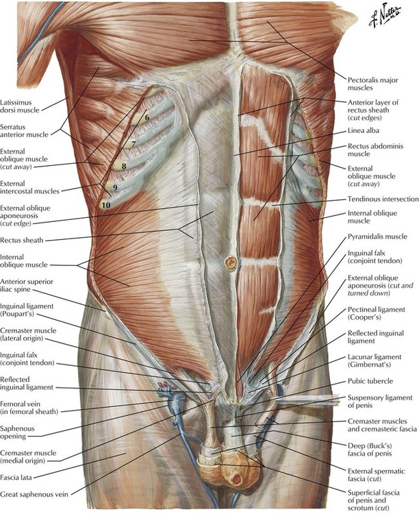
Transversus abdominis muscle internal abdominal oblique muscle rectus abdominis muscle external abdominal oblique muscle pyramidalis muscle. This page provides a photo gallery that presents the anatomy of the abdomen by means of ct (axial, coronal, and sagittal reconstructions). The abdomen (colloquially called the stomach, belly, tummy or midriff) is the part of the body between the thorax (chest) and pelvis, in humans and in other vertebrates. Sciency root words make anatomical parts harder to memorize. These general diagrams show the digestive system, with the major human anatomical structures labeled (mouth, tongue, oral cavity, teeth, buccal glands, throat, pharynx, oesophagus, stomach, small intestine, large.
anatomy test 1 - Speech Language Hearing Science 406 with Vogelman at Loyola College - StudyBlue from classconnection.s3.amazonaws.com The transversus abdominis muscle is the deepest of the abdominal muscles, lying internally to the internal abdominal obliques. The abdomen (colloquially called the stomach, belly, tummy or midriff) is the part of the body between the thorax (chest) and pelvis, in humans and in other vertebrates. Abdominal anatomy seen on ct. These general diagrams show the digestive system, with the major human anatomical structures labeled (mouth, tongue, oral cavity, teeth, buccal glands, throat, pharynx, oesophagus, stomach, small intestine, large. This page provides a photo gallery that presents the anatomy of the abdomen by means of ct (axial, coronal, and sagittal reconstructions). Abdomen, in human anatomy, the body cavity lying between the chest or thorax above and the pelvis below and from the spine in the back to the wall the abdominal organs are supported and protected by the bones of the pelvis and ribcage and are covered by the greater omentum, a fold of peritoneum. Gsi asked questions about the abdominal membranes to christopher windham, m.d. Sciency root words make anatomical parts harder to memorize.
Abdominal surface anatomy can be described when viewed from in front of the abdomen in 2 ways:
Lee moffitt cancer center & research institute in. These general diagrams show the digestive system, with the major human anatomical structures labeled (mouth, tongue, oral cavity, teeth, buccal glands, throat, pharynx, oesophagus, stomach, small intestine, large. Simple, easy notes for quick revision of important questions. Transversus abdominis muscle internal abdominal oblique muscle rectus abdominis muscle external abdominal oblique muscle pyramidalis muscle. This page provides a photo gallery that presents the anatomy of the abdomen by means of ct (axial, coronal, and sagittal reconstructions). Windham was previously a surgical oncologist in the sarcoma program of the h. Abdominal anatomy seen on ct. Abdominal wall anatomy that is clinically pertinent to the surgeon, focusing primarily on the structures of the anterior abdominal wall, will be reviewed. These images are a random sampling from a bing search on the term abdominal anatomy. click on the image (or right click) to open the source website in a new browser window. Abdominal surface anatomy can be described when viewed from in front of the abdomen in 2 ways: The xiphoid process and costal. Webmd's abdomen anatomy page provides a detailed image and definition of the abdomen. The abdomen (colloquially called the stomach, belly, tummy or midriff) is the part of the body between the thorax (chest) and pelvis, in humans and in other vertebrates.
Share :
Post a Comment
for "Abdominal Anatomy / Internal Anatomy Of Male Chest And Abdomen On White Stock Photo - Download Image Now - iStock"


Post a Comment for "Abdominal Anatomy / Internal Anatomy Of Male Chest And Abdomen On White Stock Photo - Download Image Now - iStock"